Mass Photometry
Institute of Molecular Biology and Pathology (IBPM) - Roma
Description
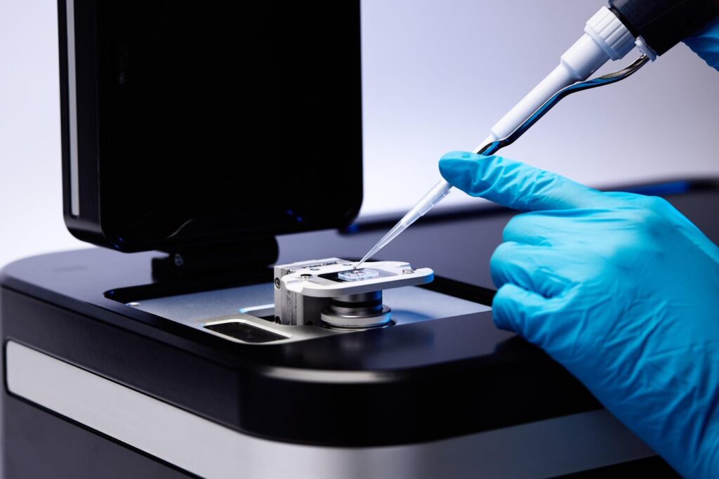
Mass photometry, an analytical technology for biomolecular characterization, measures the mass of single particles in solution. The principle behind mass photometry is simple: a single molecule in contact with a measurement surface (e.g. a coverslip), exposed to a beam of light, produces a small but measurable light scattering signal, which is directly proportional to the molecule’s mass. The mass-photometric apparatus (TwoMP, Refeyn) at IBPM-CNR (Biocrystal Facility) offers the opportunity to measure the oligomeric state of the protein sample to be crystallized. Moreover, with mass photometry is possible to perform a biophysical characterization of the biological macromolecules. In particular, it is possible to study antibody-antigen interactions, quantify small-molecule induced changes to complex formation, assess sample purity and study the thermodynamic of macromolecule complex formation.
Instruments
O.U. P.I.: Andrea Ilari
People Involved: Adele Di Matteo, Carmelinda Savino, Beatrice Vallone, Giorgio Giardina, Linda Celeste Montemiglio
Application: Biophysical characterization of protein sample: Mass Photometry
Mass photometry is an innovative technique to measure the molecular mass of biological macromolecules. Unlike other techniques, mass photometry does not infer the mass indirectly from a different physical parameter, such as the hydrodynamic radius. Indeed, the mass photometry signal measured is directly correlated with the true molecular mass, enabling measurement of the mass of molecules in the range 30 kDa to 5 MDa. Since Mass photometry measures the mass of each molecule that is in contact with the measurement surface, it is possible with this technique to detect subpopulations of protein species or detail the molecular heterogeneity – aspects of a sample that are invisible to methods that use bulk measurement. Mass photometry measurements are performed in solution and are compatible with water as well as a wide range of buffers. Measuring biomolecules in an environment that mimics the intracellular aqueous environment allows their true native behaviour to be studied. One of the main advantage of this technique is that it requires very little sample: 10 µL in a concentration range of 100 pM – 100 nM. The low concentration range allows to study the biological macromolecule at a physiologically relevant concentrations, helping to mimic the intracellular environment and observe native behaviour. Mass photometry does not require any labelling of the molecules under investigation, simplifying sample preparation and eliminating the risk of labels interfering with the native behaviour of a molecule being analysed. Mass photometry workflows are very quick, with both sample preparation and measurement together often taking just minutes. It is possible also to use this technique to capture the dynamic behaviour of biomolecules, to monitor shifts in chemical equilibria or to study the assembly/disassembly of macromolecular complexes or reveal the existence of transient intermediates or steps in the formation of protein oligomers – all phenomena that could be missed when relying on static snapshots (and/or population-averaged data). With mass photometry, the Biocrystal facility offers to the Italian scientific community the opportunity to characterize their biological samples and in particular: to measure the molecular mass of single biomolecules, oligomers, polymers, macromolecular assemblies and nanostructures (Chen, et al. 2021, England, et al. 2020, Bertosin, et al. 2021, Naftaly, et al. 2021); to quantify the oligomerisation and aggregation of biomolecules (Naftaly, et al. 2021); to characterise sample heterogeneity (Olerinyova, et al. 2021, Sonn Segev, et al. 2020); to monitor the stability of sample components (Nuber, et al. 2021); to study the effects of molecular or experimental modifications on sample integrity (Bertosin, et al. 2021); to study the biomolecular interactions, including protein-protein interactions (Higuchi, et al. 2021; Soltermann, et al. 2020) and protein-DNA interactions (Hickman, et al. 2020; Acharya, et al. 2021).