Cryogenic Electron Microscopy

CNR-IC URT Caserta

Transmission electron cryomicroscopy (cryo-EM) stands as a revolutionary tool in the field of structural biology, enabling scientists to peer into the intricate machinery of life. This cutting-edge technique has revolutionized our understanding of biological macromolecules by transforming our ability to visualize molecular structures at near-atomic resolution (Fig.1). Over the last decade, cryo-EM has found applications across a diverse array of biological systems, offering insights into the architecture of proteins, virus assembly, as well as cellular organization.

Figure 1: cryo-EM structure of key cancer signaling protein attached to the cancer drug
(Castells-Graells et al., 2023, PNAS | vol. 120 | no. 37)

Figure 2: Sample preparation by plunge-freezing 

(Murata & Wolf, 2018, BBA – General Subjects, 1862, 324–3)

At the heart of cryo-EM lies the sample vitrification, which consists on the fixation of the sample of interest within a thin layer of vitreous ice – via rapid plunge freezing. This crucial process aim to prevent the formation of damaging ice crystals, ensuring so the preservation of biological samples in their native hydrated state (Fig. 2).
Once vitrified, the specimen is then transferred to the cryo-EM microscope which operates at nitrogen temperature to maintain the sample in its frozen state. In the microscope a beam of electrons is directed at the sample, producing 2D images that are captured by a dedicated detector. Multiple images – so called micrographs – are hence collected allowing the alignment and averaging of thousands of individual single proteins to enhance the signal-to-noise ratio. Mathematical approaches, such as back-projection, are then used to reconstruct a 3D model of the specimen. This process is named Single Particle Analysis (SPA) (Fig. 3).

Figure 3: SPA-cryo EM data analysis workflow.

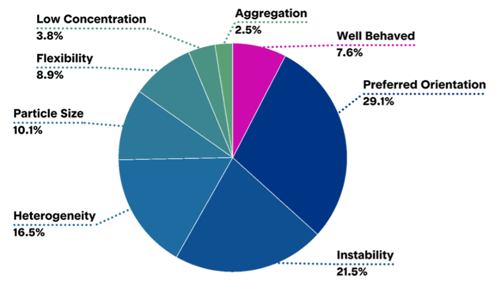
However, less than 10% of the samples appear on the grid suitable for high-resolution imaging (Fig. 4). A pre-screening step to evaluate the purity, concentration, and stability of the sample, as well as the quality of the grid and the collection settings, is therefore crucial for the complete success of the experiment as deviations in grid quality can easily result in poor icing or poor sample distribution, affecting the quality of the final images.

Figure 4: Quality of samples on the grids following the vitrification process.
(https://www.nanoimagingservices.com/blog/one-view-that-rules-them-all-and-other-vitrification-hurdles-in-cryo-em)

Screening is, by far, an expensive and time-consuming task, which might repel researchers from using cryoEM in their studies. At CNR-IC URT in Caserta, we provide the community with easy and open access cutting-edge instrumentation for screening as well as data collection opportunities in the field of SPA cryoEM.

References:
1. Cheng Y. (2018). Single-Particle Cryo-EM at Crystallographic Resolution. Cell, 171(2), 318–327.
2. Nogales E., Scheres S. H. W. (2015). Cryo-EM: A Unique Tool for the Visualization of Macromolecular Complexity. Molecular Cell, 58(4), 677–689.
3. Bai X. C., McMullan G., Scheres S. H. W. (2015). How Cryo-EM Is Revolutionizing Structural Biology. Trends in Biochemical Sciences, 40(1), 49–57.
4. Kühlbrandt W. (2014). The Resolution Revolution. Science, 343(6178), 1443–1444.
5. Subramaniam S., Kühlbrandt W., Henderson R. (2016). Cryo-EM at IUCrJ: A new era. IUCrJ, 3(Pt 1), 3–7.

Equipment:
Glacios: 200 kV accelerating voltage, field emission gun (X-FEG) source, equipped with an autoloader, ideal for cryo-EM sample optimization and data collection.
Vitrobot Mark IV System: a state-of-the-art instrument widely used for sample preparation. It operates within a controlled environment, maintaining consistent temperature and humidity levels.
Data storage
We use 580TB HD for data storage
High-performance computing cluster
A computing cluster allowing efficient data processing using state-of-the-art data processing software such as Relion or CryoSPARC.